

Chances are good that if you’re involved with an injury or chronic pain, or you have a doctor trying to see very fine details of your body’s interior, you’ve had an MRI. MRI most assuredly stands for an advanced and certainly among the priciest diagnostic tool, which pictures, and very sharply at that, not just the hard things like your bones but also all the soft tissues in your body. And ostensibly, MRI imaging Los Angeles does this totally without using radiation.
What is MRI?
Detailed internal images of the body are made with the help of radio waves and powerful magnets. These images result from examining the reactions of the body’s hydrogen atoms to radio waves and strong magnetic fields.
There is water in huge quantities inside your body. Inside the water molecule, there is a hydrogen atom and it is a weak magnet. MRI machines sense these types of atoms and these atoms are used to make images with great accuracy. That’s the reason why muscle, ligament, brain, spinal cord, and internal organs are shown by MRI in Los Angeles this much clearly.
How does an MRI machine work?
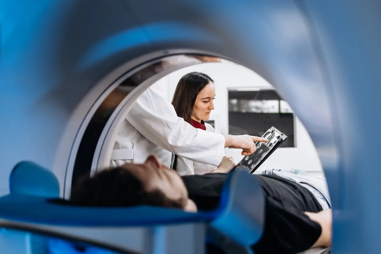
An MRI imaging machine is a large, tubular machine inside a massive magnet. As you lie on the bed and roll into the machine, you roll into a very strong magnetic field. The magnetic field aligns your body’s hydrogen protons with the direction of the small compass dipoles.
Then the machine sends out a burst of radio waves through your body. Radio waves turn the hydrogen protons in and out of alignment for a split second. Once the burst has passed, the protons relax back into the magnetic field. When they move back to their original position, they release energy. The MRI machine detects the energy and uses it to construct cross-sectional images.
How the different reflection of signals from your body as signals differs with what kind of tissue it is. Fat tissue, muscle tissue, and fluid tissue, for example. Because MRI scanning can detect differences between tissues that other scanning techniques cannot.
What kind of images does MRI produce, apart from X-ray ones?

MRI Los Angeles pictures generally arrive in slices. They are sort of thin cross-sections of your body, similar to cutting a loaf of bread. Radiologists can view the slices from any direction—they can view the front and back, top and bottom, side to side. Multi-direction viewing provides a very realistic sense of what is happening in your body.
Contrast agents can be used to improve MRI. They are drugs, maybe intravenous drugs, and they are prescribed only to make certain vascular structures or tissues brighter so that they will be easier to see. MRI with contrast, for example, is used in imaging tumor, inflammation or disease of the arteries.
What is the main clinical application of MRI?
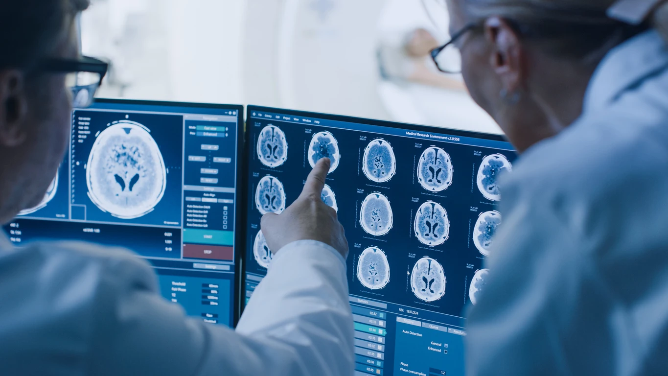
MRI scanner is used in the diagnosis of the majority of medical illnesses. MRI is most suited to image soft tissue and thus has to do brain, spinal cord, joint, and visceral organ imaging. MRI is most used by the department of neurology in its diagnostic process resulting in diagnosis of brain tumor, stroke, multiple sclerosis, and spinal disc disease.
Orthopedic MRI is used in joint trauma imaging, cartilage and ligament ruptures. It is used in abdominal imaging through disease of the bowel and renal masses, and for liver disease diagnosis. It is used mainly in cardiovascular imaging to create a picture of the heart anatomy and a picture of blood flow.
MRI is a non-invasive test and the patient does not need to have to open up their body for a diagnosis to be obtained. It is particularly effective for tracking the long-term progression of slow-moving diseases and their effects in postoperative patients.
How does MRI differ from other imaging scans l?
The main advantage of MRI scans is that they don’t use radiationl. X-rays and CT scans amount to ionizing radiation exposure in poisonous dosages where it is toxic. MRI utilizes magnetic fields and radio waves sa after substance to inject into standard protocol, particularly to more delicate or child patients.
MRI makes pictures that are more tissue-sensitive than CT or X-ray. CT’s bone image and acute trauma is good, but MRI is disease-sensitive to organ, muscle and nerve.
MRI is also most likely to be slower and less expensive than CT or X-ray. It’s also on this basis in part that doctors make a decision about how to type of scan to order depending on what medical question it’s attempting to answer.
My MRI patient experience?
You won’t experience anything for the majority of patients who have an MRI test. You might lie down or sit on a level bed, and the bed will be moved into the doughnut-shaped machine. The scan itself takes only 20 minutes to as long as an hour or more depending on where in your body you’re getting scanned.
The scanner is beeeping/tapping while it’s taking the pictures. Earphones/headphones are also provided to the patient so the sound is muted. Walk softly as possible to get the scan so one has awesome pictures. Sedation is employed in some cases, i.e., agitated patients and children.
One of the limitations of MRI is it’s not for everyone. There are pacemaker patients, metal implant patients, or claustrophobic patients who will need to have other tests completed on them. That is why safety screens always come first.
Is MRI suitable for preventative care?
Yes. Pre-symptomatic MRI screening also is hip as a disease prevention measure prior to disease. Whole-body MRI, for example, now is being marketed as a means of identifying precursors to cancer, vascular disease, or neurological injury. Not yet standard of care anywhere, whole-body MRI screening is being demanded by patients who desire an active life.
Other than that, MRI is used in patient follow-up with chronic disease. For example, a patient with multiple sclerosis would undergo follow-up MRIs in long-term observation of lesion change in the brain or spinal cord. It can be used in treatment planning and possible future development of the disease.
What does the future hold for MRI technology?

The future of MRI is not bleak. Scientists are working on shorter scan time, better images, and open scanner design to make it less invasive. AI technology also proves useful with better image interpretation, better scan quality, and avoidance of unnecessary scanning.
One of the newer trends, though, is towards mobile MRI units. Even the compact ones can be fitted into emergency rooms, ICUs, and even rural villages where the bigger ones would be a waste.
The second of the new modalities is called functional MRI or fMRI. It photographs brain activity by scanning for changes in blood flow. It is applied to the study of neuroscience, and one hopes someday to psychiatric disease or head injury treatment.
Conclusion
MRI scanning is man’s most precise medical diagnostic device for medicine, surgery-free and genuine windows into human bodies’ inner mechanisms. MRI continues to reveal new horizons for medical practice, from observing brain disease to evaluating joint trauma—and its potential is barely less exciting.
Share This Story, Choose Your Platform!
More Posts You May Find Interesting
Once upon a time, to go for a cancer screening meant that you had to feel symptoms and visit the doctor, or be a
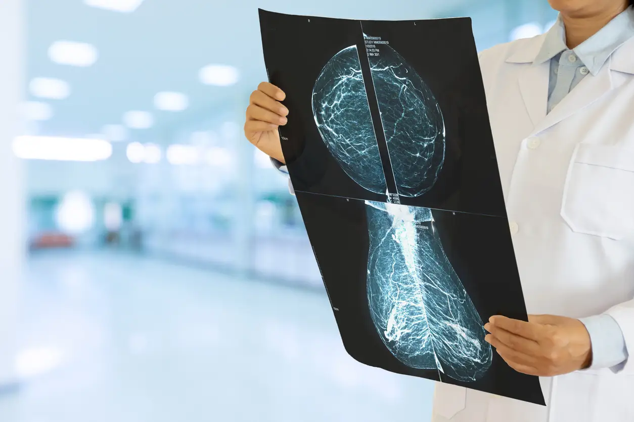
Although breast cancer remains as one of the major cancer diseases in women worldwide. Early detection is one of the best ways to improve

If you are scheduled for an MRI, there are a few things that you need to do in advance for special preparation. A single


